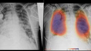
UC San Diego Health radiologists and other physicians are now using artificial intelligence to rapidly analyze lung imaging to detect for pneumonia -- including cases impacting COVID-19 patients, the medical center announced Tuesday.
For most patients who have died of COVID-19, the ultimate cause of death was pneumonia, a condition in which inflammation and fluid buildup make it difficult to breathe.
Severe pneumonia often requires lengthy hospital stays in intensive care units and assistance breathing with ventilators -- medical devices now in high demand in some cities grappling with a surge of COVID-19 cases.
To quickly detect pneumonia -- and therefore better distinguish between COVID-19 patients likely to need more supportive care in the hospital and those who could be followed closely at home -- researchers are augmenting lung imaging analysis in a clinical research study enabled by Amazon Web Services.
The new AI capability has so far provided UC San Diego Health physicians with unique insights into more than 2,000 images, researchers say. In one case, a patient in the emergency department who did not have any symptoms of COVID-19 underwent a chest X-ray for other reasons.
Yet the AI readout of the X-ray indicated signs of early pneumonia, which was later confirmed by a radiologist. As a result, the patient was tested for COVID-19 and found to be positive for the illness.
"We would not have had reason to treat that patient as a suspected COVID-19 case or test for it, if it weren't for the AI,'' said Dr. Christopher Longhurst, chief information officer and associate chief medical officer for UC San Diego Health. "While still investigational, the system is already affecting clinical management of patients."
Local
The new capability got its start several months ago when Dr. Albert Hsiao, associate professor of radiology at UC San Diego School of Medicine, and his team developed a machine learning algorithm that allows radiologists to use AI to enhance their own abilities to spot pneumonia on chest X-rays.
"Pneumonia can be subtle, especially if it's not your average bacterial pneumonia, and if we could identify those patients early, before you can even detect it with a stethoscope, we might be better positioned to treat those at highest risk for severe disease and death," Hsiao said.
More recently, Hsiao's team applied this AI approach to 10 chest X-rays, published in medical journals, from five patients treated in China and the United States for COVID-19.
The algorithm consistently localized areas of pneumonia, despite the fact that the images were taken at several different hospitals, and varied considerably in technique, contrast and resolution.
Hsiao's AI method has been deployed across UC San Diego Health in a clinical research study that allows any physician or radiologist to get an initial estimate regarding a patient's likelihood of having pneumonia within minutes, at point-of-care.
According to Hsiao, chest X-rays are cheaper, the equipment is more portable and easier to clean, and results are returned more quickly than many other diagnostics.
To be clear, UC San Diego Health experts emphasize they are not diagnosing COVID-19 itself by lung imaging. Pneumonia can be caused by several different types of bacteria and viruses.
In addition, the use of Hsiao's AI algorithm is still considered investigational. Although these images are available for use by clinicians, patient care is still guided by formal interpretation from human radiologists.
Next, the UC San Diego Health team hopes to expand the AI-powered study for detecting pneumonia to the UC system's four other academic medical centers.


